Index
Long QT Syndrome
What Is Long QT Syndrome?
Long QT syndrome (LQTS) is a condition in which repolarization of the heart after a heartbeat is affected.
It results in an increased risk of an irregular heartbeat which can result in fainting, drowning, seizures, or sudden death.
These episodes can be triggered by exercise or stress.
Some rare forms of LQTS are associated with other symptoms and signs including deafness and periods of muscle weakness.
These episodes can be triggered by exercise or stress.
Some rare forms of LQTS are associated with other symptoms and signs including deafness and periods of muscle weakness.
What Causes Long QT Syndrome?
There are several subtypes of long QT syndrome. These can be broadly split into those caused by genetic mutations which
those affected are born with, carry throughout their lives, and can pass on to their children (inherited or congenital long QT syndrome),
and those caused by other factors which cannot be passed on and are often reversible (acquired long QT syndrome).
What are the Signs & Symptoms of Long QT Syndrome?
Long QT syndrome have no signs or symptoms. When symptoms occur, they are generally caused by abnormal heart rhythms (arrhythmias),
most commonly a form of ventricular tachycardia called Torsades de pointes (TdP). If the arrhythmia reverts to a normal rhythm spontaneously
the affected person may experience lightheadedness (known as Presyncope) or faint which may be preceded by a fluttering sensation in the chest.
If the arrhythmia continues, the affected person may experience a cardiac arrest, which if untreated may lead to sudden death. Those with LQTS may also experience seizure-like activity (non-epileptic seizure) as a result of reduced blood flow to the brain during an arrhythmia.
Epilepsy is also associated with certain types of long QT syndrome.
HEART CONDITIONS Diseases and Treated FAQ's
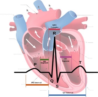
Diagnosing long QT syndrome is challenging. The QT interval is highly variable among both those who are healthy and those who have LQTS.
This leads to overlap between the QT intervals of those with LQTS and those without – 2.5% of those with LQTS have a normal QT interval while 10–15% of the healthy population has a prolonged QT.
- Other factors beyond the QT interval should be considered when making a diagnosis, some of which have been incorporated into scoring systems.
- Electrocardiogram,
- Schwartz score.
The first operations to treat coarctation were carried out by Clarence Crafoord in Sweden in 1944.
Some patients found to have coarctation, treatment is conservative if asymptomatic, but may require surgical resection of the narrow segment if there is arterial hypertension.
Treatment options for coarctation of the aorta depend on your age at the time of diagnosis and the severity of your condition.
Other heart defects might be repaired at the same time as aortic coarctation. Treatment approaches usually consist of surgery or a procedure called balloon angioplasty or stent placement. In some cases angioplasty can be performed to dilate the narrowed artery, with or without the placement of a stent graft.
Risk factors Coarctation of the aorta often occurs along with other congenital heart defects,
although doctors don't know what causes multiple heart defects to form together. The condition is more common in males than in females.
You or your child may be more likely to have aortic coarctation if certain heart conditions exist, including:
- Bicuspid Aortic Valve
The aortic valve separates the lower left chamber (left ventricle) of the heart from the aorta. A bicuspid aortic valve has two flaps (cusps) instead of the usual three. Many people with coarctation of the aorta have a bicuspid aortic valve.
- Patent Ductus Arteriosus
Before birth, the ductus arteriosus is a blood vessel connecting the left pulmonary artery to the aorta — allowing blood to bypass the after birth, the ductus arteriosus usually closes. If it remains open, it's called a patent ductus arteriosus.
- Holes in the Wall (Between the left and right sides of the heart.)
You may have a hole in the wall (septum) between the upper chambers of the heart (atrial septal defect) or the lower chambers of the heart (ventricular septal defect) when you're born. This causes oxygen-rich blood from the left side of the heart to mix with oxygen-poor blood in the right side of the heart.
- Aortic Valve Stenosis
This is a narrowing of the valve that separates the left ventricle of the heart from the aorta (aortic valve). This means your heart has to pump harder to get adequate blood flow to your body. Over time, this can cause your heart muscle to thicken and lead to symptoms such as chest pain, fainting spells and breathlessness, or heart failure.
- Aortic Valve Regurgitation
This occurs when the aortic valve doesn't close tightly, causing blood to leak backward into the left ventricle.
- Mitral Valve Stenosis
This is a narrowing of the valve (mitral valve) between the upper left heart chamber (left atrium) and the left ventricle that lets blood flow through the left side of your heart. In this condition, blood may back up into your lungs, causing shortness of breath or lung congestion. Like aortic valve stenosis, this condition can also lead to heart failure.
- Mitral valve regurgitation
This occurs when the mitral valve doesn't close tightly, causing blood to leak backward into the left atrium.