Index
Double Outlet of the Right Ventricle
What Is Double Outlet of the Right Ventricle?
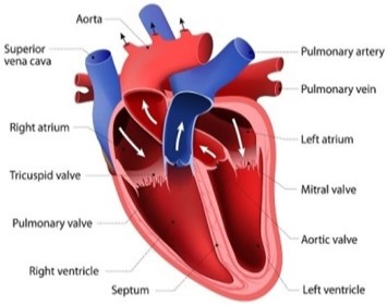
Double outlet right ventricle (DORV) is a form of congenital heart disease where both of the great arteries
connect (in whole or in part) to the right ventricle (RV). In some cases it is found that this occurs on the left side of
the heart rather than the right side.
What Causes Double Outlet of the Right Ventricle?
Double Outlet of the Right Ventricle is due to an error in the way the heart forms early in pregnancy.
Why this happens is unknown. It is not caused by anything a mother did or did not do during her pregnancy.
Doctors and scientists have not yet found a way to prevent.
What are the Signs & Symptoms of Double Outlet of the Right Ventricle?
- A heart murmur (an extra, unusual heart sound).
- Breathing problems, including difficulty breathing,
- Shortness of breath, or rapid breathing.
- Difficulty eating and/or gaining weight.
- Blueness of the lips, nails, and/or skin (Cyanosis.)
- Sweating and Fatigue.
- Breathing problems, including difficulty breathing,
- Shortness of breath, or rapid breathing.
- Difficulty eating and/or gaining weight.
- Blueness of the lips, nails, and/or skin (Cyanosis.)
- Sweating and Fatigue.
HEART CONDITIONS Diseases and Treated FAQ's
- Pulse Oximetry
Measurement of the oxygen in blood using a light at a fingertip or toe.
- Echocardiography
This is the most important test. It shows the movement of blood through the valve.
- Transesophageal Echocardiography
This is a heart ultrasound done from the esophagus that can give better pictures of the aortic valve.
- Electrocardiogram
This is done to check the heart’s electrical rhythm.
- Chest X-ray
These help view the heart anatomy and lungs.
- Cardiac CT or MRI
These are done if more detail is needed about the valve, heart, or aorta.
Intraventricular Repair
- This surgery uses a patch to make a tunnel from the VSD to the aorta. Then, when the heart contracts, the oxygen-rich blood in the left ventricle flows through the VSD into the aorta. After the repair, there is no longer mixing of oxygen-rich and oxygen-poor blood in the right ventricle.
- Arterial Switch
The arterial switch repair moves the aorta from the right ventricle to the left ventricle. The VSD is closed to keep oxygen-rich and oxygen-poor blood from mixing in the ventricles.
- Single Ventricle Pathway
In some cases many surgical steps (called the single ventricle pathway) are needed to balance blood flow in a baby with DORV:
- At Age of 2 weeks or Younger
A Blalock-Taussig (BT) shunt redirects some of the left ventricle's output from the body to the lungs.
– OR – If the blood flow to the lungs is too high, as can happen with a large VSD, a band around the pulmonary artery lessens the flow to prevent damage.
- At age 4 ‒ 6 Months
The Glenn procedure allows blood returning from the upper part of the body to flow directly to the lungs. The BT shunt is removed at the same time.
- At age 1.5 ‒ 3 Years
The Fontan procedure channels blood from the lower half of the body to the lungs so the heart pumps oxygen-rich blood to the body only. Blood returning from the body flows to the lungs before passing through the heart.
In rare cases, surgical repair is not possible, and a heart transplant may be recommended.