Index
Ebstein's Anomaly
What Is Ebstein’s Anomaly?
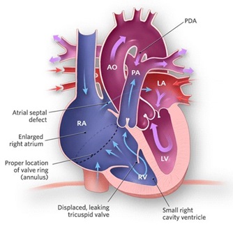
Ebstein's Anomaly is a congenital heart defect in which the septal and posterior leaflets of the
tricuspid valve are displaced towards the apex of the right ventricle of the heart.
It is classified as a critical congenital heart defect accounting for <1% of all congenital heart defects presenting
in ≈1 per 200,000 live births.
What Causes Ebstein’s Anomaly?
Until today, Doctors do not know exactly why a baby's heart develops Ebstein anomaly during pregnancy.
But it is not caused by anything a mother does or does not do during her pregnancy.
What are the Signs & Symptoms of Ebstein’s Anomaly?
- The annulus of the valve is still in the normal position. The valve leaflets, however, are to a varying degree,
attached to the walls and septum of the right ventricle.
A subsequent "atrialization" of a portion of the morphologic right ventricle (which is then contiguous with the right atrium) is seen.
This causes the right atrium to be large and the anatomic right ventricle to be small in size.
- S3 heart sound.
- S4 heart sound.
- Triple or quadruple gallop due to widely split S1 and S2 sounds plus a loud S3 and/or S4.
- Systolic murmur of tricuspid regurgitation = Holosystolic or early systolic murmur along the lower left sternal border depending on the severity of the regurgitation.
- Right atrial hypertrophy.
- Right ventricular conduction defects.
- Wolff-Parkinson-White syndrome often accompanies.
HEART CONDITIONS Diseases and Treated FAQ's
- Echocardiogram
The best test to confirm Ebstein anomaly is an echocardiogram (ultrasound of the heart).
- Pulse Oximetry
Measurement of the oxygen in blood using a light at a fingertip or toe.
- Echocardiography
This is the most important test. It shows the movement of blood through the valve.
- Transesophageal Echocardiography
This is a heart ultrasound done from the esophagus that can give better pictures of the aortic valve.
- Electrocardiogram
This is done to check the heart’s electrical rhythm.
- Chest X-ray
These help view the heart anatomy and lungs.
- Cardiac CT or MRI
These are done if more detail is needed about the valve, heart, or aorta.
- Exercise test
Some appropriate exercises measure the power of heart and their condition.
- If the defect was found before birth, the delivery team will be ready to provide intensive care immediately in case the newborn is not doing well.
But most newborns with the anomaly do not need immediate treatment. Doctors will monitor them closely to watch for changes and provide quick treatment if needed.
- Some children with Ebstein anomaly do not need treatment. When treatment is needed, the most common types used are:
- Oxygen,
- Surgery,
- Medicines,
- Electrophysiology Study.
- Some children with Ebstein anomaly do not need treatment. When treatment is needed, the most common types used are:
- Oxygen,
- Surgery,
- Medicines,
- Electrophysiology Study.
An enlargement of the aorta may occur; an increased risk of abnormality is seen in babies of women taking lithium
during the first trimester of pregnancy (though some have questioned this) and in those with Wolff-Parkinson-White syndrome.