Index
Arterial Trunk
What Is Arterial Trunk?
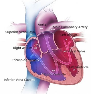
Also called Persistent Truncus Arteriosus is a solitary trunk that exits the heart through a
common ventricle-arterial junction and supplies directly the systemic, pulmonary, and coronary arterial pathways.
This is a rare form of congenital heart disease that presents at birth. In this condition, the embryological structure known as the truncus arteriosus fails to properly-divide into the pulmonary trunk and aorta. This results in one arterial trunk arising from the heart and providing mixed blood to the coronary arteries, pulmonary arteries, and systemic circulation.
This is a rare form of congenital heart disease that presents at birth. In this condition, the embryological structure known as the truncus arteriosus fails to properly-divide into the pulmonary trunk and aorta. This results in one arterial trunk arising from the heart and providing mixed blood to the coronary arteries, pulmonary arteries, and systemic circulation.
What Causes Arterial Trunk?
Most of the time, this defect occurs spontaneously.
Genetic disorders, and teratogens (viruses, metabolic imbalance, and industrial or pharmacological agents) have been associated as possible causes. Up to 50% (varies in studies) of cases are associated with chromosome 22q11 deletions (DiGeorge Syndrome). The neural crest, specifically a population known as the cardiac neural crest, directly contributes to the aorticopulmonary septum.
Genetic disorders, and teratogens (viruses, metabolic imbalance, and industrial or pharmacological agents) have been associated as possible causes. Up to 50% (varies in studies) of cases are associated with chromosome 22q11 deletions (DiGeorge Syndrome). The neural crest, specifically a population known as the cardiac neural crest, directly contributes to the aorticopulmonary septum.
Microablation of the cardiac neural crest in developing chick embryos and genetic anomalies affecting this
population of cells in rodents results in persistent truncus arteriosus.
What are the Signs & Symptoms of Arterial Trunk?
- Symptoms of truncus arteriosus include rapid breathing (tachypnea), lethargy, a bluish tint to the skin (cyanosis), poor feeding,
difficulty breathing (dyspnea), and broadening of the fingertips (clubbing).
- Other symptoms may include abnormal accumulations of fluid in the face, arms, and/or legs (edema), an abnormally rapid heartbeat, slow weight gain, failure to thrive, recurrent respiratory infections, poor physical development, and/or growth delays.
- The symptoms of truncus arteriosus are similar to those of a severe ventricular septal defect (VSD), a relatively common form of congenital heart disease. In VSD, the wall (septum) separating the two ventricles is incompletely formed before birth. This can result in inefficient distribution of oxygen to the various tissues of the body (hypoxia) and various degrees of congestive heart failure.
HEART CONDITIONS Diseases and Treated FAQ's
- Cyanosis presents at birth,
- Heart failure may occur within weeks,
- Systolic ejection murmur is heard at the left sternal border,
- Widened pulse pressure,
- Bounding arterial pulses,
- Loud second heart sound,
- Biventricular hypertrophy,
- Cardiomegaly,
- Increased pulmonary vascularity,
- Hypocalcemia (if associated with DiGeorge syndrome).
- Treatment is with neonatal surgical repair, with the objective of restoring a normal pattern of blood flow.
The surgery is open heart, and the patient will be placed on cardiopulmonary bypass to allow the surgeon to work on a still heart.
- The heart is opened and the ventricular septal defect is closed with a patch. The pulmonary arteries are then detached from the common artery (truncus arteriosus) and connected to the right ventricle using a tube (a conduit or tunnel).
- The common artery, now separated from the pulmonary circulation, functions as the aorta with the truncal valve operating as the aortic valve.
- Most babies survive this surgical repair but, may require further surgery as they grow up. For example, the conduit does not grow with the child and may need to be replaced as the child grows.